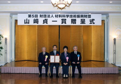
The 5th (2005) Yamazaki-Teiichi Prize Winner Biological Science and Technology
Structural and Functional Study of Membrane Proteins
| Winner | ||
|---|---|---|
| Yoshinori Fujiyoshi | ||
| Affiliation at the time of the award | ||
| Professor, EMGroup, Department of Biophysics, Faculty of Science, Kyoto University |
Reason for award
In X-ray crystallography, the method conventionally used to analyze protein structure, large 3D crystals have to be grown of the proteins under investigation. This is very difficult to accomplish with membrane proteins. In the cryo-electron microscope developed by Dr. Fujiyoshi the specimen is cooled down to the temperature of liquid helium, which substantially reduces the effects of radiation damage. This ingenious innovation made it possible to use electron microscopy to solve the 3D structures of channels, receptors and other membrane proteins.
In conjunction with Dr. Ueda, Dr. Fujiyoshi proved that molecular structures can be directly observed in the electron microscope. Subsequently, he has developed a cryo-electron microscope that keeps specimens at liquid helium temperature, making it possible to take high-resolution images even of specimens that are easily damaged by the electron beam.
Dr. Fujiyoshi has applied his newly developed microscope to the analysis of membrane proteins, which play crucial roles in signal transduction mechanisms in cells. He has elucidated the structures of a light-harvesting antenna protein, bacteriorhodopsin, several aquaporin water channels, the nicotinic acetylcholine receptor, and a number of other important membrane proteins. Today, cryo-electron microscopes with a helium-cooled specimen stage are not only used by Dr. Fujiyoshi, but make contributions to the research of many groups across the globe, including research groups in the US and Europe.
The results obtained by Dr. Fujiyoshi and his colleagues on the structures of integral membrane proteins have had a great impact on our understanding of their functions. Dr. Fujiyoshi has vigorously worked towards elucidating the molecular structure and the physiological functions of the many types of water channels present in humans. He has also helped to clarify the overall structure of sodium channels.
By analyzing the structure of the nicotinic acetylcholine receptor, Dr. Fujiyoshi and his colleagues have contributed tremendously to our understanding of the structure of gated ion channels and the mechanism by which neurotransmitters mediate signal transduction. This research also contributed to our understanding of the effects of alcohol and anaesthetics. For his work Dr. Fujiyoshi has received great attention in the medical field, and it garnered him high praise worldwide.
As described above, using the cryo-electron microscope he developed, Dr. Fujiyoshi has produced results on the very highest level. His analysis of the molecular structures of many membrane proteins of the central nervous system as well as of membrane proteins that play major roles in other biological functions deeply influenced the thinking in the life sciences. As all of his results are also practical, directly linking the elucidation of biological functions to medical care, Dr. Fujiyoshi has been awarded this prize.
Background of research and development
When Dr. Max Perutz introduced the isomorphous replacement method to phase intensity data sets, X-ray crystallography became the central method for the analysis of protein structure. In 1985, Deisenhofer et al. used this method to solve the first structure of a membrane protein to atomic resolution. However, 10 years earlier, in 1975, Drs. Henderson and Unwin at the MRC Laboratory of Molecular Biology in the UK developed an alternative approach, electron crystallography, and used it to determine the structure of bacteriorhodopsin to a resolution of 7Å. Dr. Henderson and his colleagues then spent 15 years to develop electron crystallography to a level that allowed them to solve the structure of bacteriorhodopsin to near-atomic resolution. The resolution perpendicular to the membrane plane remained however limited for reasons such as the limited tilt angles at which data can be collected in electron microscopy (resulting in the missing cone problem) and damage to the specimen caused by the electron beam.
Achievements
The prize winner and others used chlorinated copper phthalocyanine as a test specimen to prove that electron microscopy can provide atomic resolution images of organic molecules (Chemica Scripta., 14, 47-61(1978)).
Obtaining an image with the best focus condition was however a tedious task that required a huge number of micrographs to be taken, because the molecular features seen in the images were strongly affected by even minute changes in focus conditions. Moreover, exposure with the electron beam during focusing caused damage to the specimen, making it difficult to accurately adjust the focus. To overcome this problem, the prize winner and co-workers developed a minimum dose system (MDS) that eliminated the needless pre-exposure of the imaging area during focusing.
Use of this MDS made it possible to record atomic resolution images even of organic semiconductors, such as Ag-TCNQ complexes (Nature, 285, 95-97(1980)). For the structure determination of biological macromolecules, the permissible electron dose is however extremely small. The rates with which diffraction intensities fade as a result of beam damage were measured as a function of the accumulated electron dose.
These measurements revealed that cooling the specimen to very low temperatures reduced beam-induced damage by orders of magnitude compared to exposures at room temperature. In 1986, the prize winner and co-workers therefore developed the first generation of a cryo-electron microscope that was equipped with a superfluid helium stage. Working with JEOL Ltd., the prize winner continued to improve the helium technology, leading to second- and third-generation cryo-electron microscopes (for details, see review article: Adv. Biophys., 35, 25-80 (1998)). Cryo-electron microscopes of the third and fourth-generation came to be widely used both domestically and overseas, and the recently completed fifth-generation instruments have now also been optimized for the use in single-particle electron microscopy studies.
Electron crystallography, capable of elucidating the structure of membrane proteins in lipid membranes, has become a powerful method. To date, all four types of membrane proteins analysed by electron crystallography to high resolution were imaged with cryo-electron microscopes that were equipped with the helium stage developed by the prize winner.
The analysed membrane proteins are a light-harvesting antenna protein (Nature, 367, 614-621 (1994)), bacteriorhodopsin (Nature, 389, 206-211 (1997)), a number of water channels including aquaporin-1 (Nature, 387, 624-627 (1997)), (Nature, 407, 599-605 (2000)), and the nicotinic acetylcholine receptor (Nature, 423, 949-955 (2003)). Aquaporin-1 is of particular interest, because it was the first structure obtained of a human membrane protein. The structure analysis revealed that the water channel adopts a novel and unusual fold. It also provided answers to puzzling questions, such as how the protein can be such a highly selective and efficient water channel, permitting the passage of 2 billion water molecules per second, while being completely impermeable to ions and protons. In collaboration with Dr. Unwin, the prize winner and Dr.
Miyazawa also analysed the structure of the nicotinic acetylcholine receptor and elucidated how ligand binding causes gating (opening and closing) of this ion channel. Recently, single-particle analysis is attracting attention, because this technique makes it possible to obtain 3D structures even when proteins do not crystallize. By applying single-particle analysis to images taken with a cryo-electron microscope equipped with a helium stage, the structures of a voltage-sensitive Na+-channel (Nature, 409, 1047-1051 (2001)) and of the IP3 receptor were analyzed (J. Mol. Biol. 336, 155-164 (2003)).
Meaning of the achievements
While originally only used in Japan, the cryo-electron microscopes developed by Dr. Fujiyoshi, the prize winner and his colleagues are now used for structural studies around the world. Together with technical developments for high-resolution analysis of membrane protein structure, these unique instruments continue to contribute to the elucidation of the 3D structure and physiology of membrane channels and receptors. Such studies are not only of fundamental importance in medicine and biology, but are also expected to expand the field of structural physiology as they will guide drug discovery.

