The 10th (2010) Yamazaki-Teiichi Prize Winner Measurement Science and Technology
Development of high-speed atomic force microscope and its application to dynamic observation of biomolecules
| Winner | ||
|---|---|---|
| Toshio Ando | ||
| History | ||
| Mar. 1980 | Doctor of Science, Completed doctoral course at Graduate School of Science and Engineering, Waseda University | |
| Apr. 1980 | Postdoctoral Fellow of University of California, San Francisco | |
| Jan. 1983 | Research Associate of University of California, San Francisco | |
| Mar. 1986 | Lecturer of Faculty of Science, Kanazawa University | |
| Aug. 1992 | Associate Professor of Faculty of Science, Kanazawa University | |
| Jul. 1996 | Professor of Faculty of Science, Kanazawa University | |
| Apr. 2009 | Professor of School of Mathematics and Physics, College of Science and Engineering, Kanazawa University | |
| Present | ||
Reason for award
To understand the relationship between structure and function of biomolecules as well as their functional mechanism, direct in-situ observation of the dynamically functioning biomolecules is necessary. To meet this demand, Toshio Ando developed a high-speed scanning microscope based on atomic force.
The time required to generate an image by a scanning probe microscope increases by the increment of the number of pixels which affects the image quality. There is a trade-off between the image quality and the image generation speed, and hence, there were limits in the image quality and imaging speed. Ando has overcome the limits and made it possible to directly observe movement of biomolecules in aqueous solutions.
To increase the imaging rate while maintaining the image quality, the sample height information at XY positions is acquired while the sample stage is accurately and quickly driven in the X, Y and Z directions with piezo actuators. When a micro probe approaches the sample surface, an atomic force is exerted between them. A minute displacement of the probe caused by the atomic force is optically detected while the feedback control system operates to drive the Z-piezo so that an appropriate magnitude of atomic force is always exerted. The X and Y-scanners move the sample two-dimensionally at a regular interval. To ensure the high image quality and scan speed, the two-dimensional scan should be quickly carried out while unwanted vibrations should be suppressed. To increase the speed of feedback scanning in the Z-direction, methods of active vibration damping and nonlinear control were used while the dynamic property of each element contained in the control system was improved. As a result, 10 to 25 images can be acquired per second, which is 300 to 750 times faster than the conventional image acquisition that takes about at least 30 seconds.
The achievement resulted in many papers and patents. The instrument has been manufactured by a company and used by researchers in the world.
As an example in the application studies, the imaging study on the molecular behavior of a membrane protein bacteriorhodopsin in response to light was reported in NATURE Nanotechnology.
The present achievement greatly contributes to the progress of biological science and therefore is suitable for the Yamazaki-Teiichi Prize in Measurement Science and Technology.
Background of research and development
For understanding the mechanism of protein function, it is the most direct approach to directly and simultaneously observe the structure and dynamics of the functioning molecules at high resolution. There are techniques to observe either the structure or dynamics of proteins. However, techniques to simultaneously observe them have not existed. To overcome this long-standing technological limit prevailing throughout life science, Ando started his research for increasing the imaging rate of atomic force microscopy (AFM), which was more than 15 years ago. Paul Hansma of UCSB in the US also started his research for speeding up AFM around the same time. However, both the researchers did not know each other’s research commencement at that time and knew about it much later.
Achievements
In AFM, a probe tip attached to the free end of a soft lever (cantilever) is brought closer to a sample surface or in contact with it. The force exerted between the tip and sample is measured by detecting a mechanical change (in deflection, amplitude, phase or resonant frequency) of the cantilever (Fig. 1). The distance between the probe and the sample in the Z direction is adjusted by feedback control to maintain the mechanical change constant. The surface structure of the sample is observed by performing the measurement and feedback-controlled scan over the whole specimen. Even a sample in liquids can be observed with a spatial resolution of 2 to 3 nm. However, since the measurement and feedback-controlled scan are repeated for every point of the sample surface while the relative horizontal position of the probe and the sample is being changed, it usually takes several minutes to acquire an image.
There are three key points for speeding up AFM. The first point is to speed up the feedback control, the second point is to suppress unnecessary vibrations generated when a sample stage scanner is quickly moved, and the third point is to satisfy both high speed and low invasiveness.

Fig. 1: Schematic showing walking myosin V (Lower left) and the principle of AFM acquiring the image of a sample while touching every point of the sample surface with the cantilever probe. (1) Cantilever, (2) Substrate.
To speed up the feedback control, the response speed of all the component devices included in AFM needs to be increased. So, Ando developed a microcantilever with a high resonant frequency, an optical-beam deflection detector applicable to the microcantilever, a high speed scanner, a fast amplitude detector, a high speed piezo driver, a high speed sensor, and others. As a vibration damping technique, he developed a method in which an LRC circuit simulating the scanner’s vibration property is utilized. To satisfy both high speed and low invasiveness, he developed a non-linear feedback control method. Many of these technology developments were applied for domestic and international patents, and some of them have already been issued.
All the developments resulted in the world highest performance of high-speed AFM. The feedback bandwidth exceeds 100 kHz, and consequently, an image can be acquired in 30-100ms under the condition of a scan area of 250 × 250 nm2 and 100 scan lines. Importantly, high-speed imaging can be carried out without disturbing the weak interaction between proteins. Although the Paul Hansma's group attained an imaging rate of 0.5 second/frame, they stopped the development and shifted to research on bone imaging. Mervin Miles' group in England followed the two groups and achieved an imaging rate exceeding the video rate. However, feedback scan is not used therein, and hence, dynamic observation of biological samples is infeasible.
To demonstrate that high-speed AFM observation is effective in protein research, Ando performed dynamic imaging of several protein systems. For example, he succeeded in filming high-resolution movies of motor protein myosin V walking along actin filaments (Nature, 2010) (Fig. 2) and light-driven proton pump bacteriorhodopsin in response to light (Nature Nanotechnology, 2010). The molecular movies not only provide ‘visual evidence’ for previously known facts but also reveal molecular behaviors that had been unknown. In the case of myosin V, the observation of the structure and dynamics revealed much more details than ever before regarding how the molecule generates force and how the unidirectional walking is made possible.
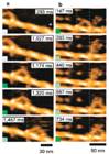
Fig. 2: Images of walking myosin V captured by HS-AFM. (a) When particles as moderate obstacles to the advance are absent on the substrate, the intermediate process during stepping is invisible as the stepping is too fast to capture. (b) When such particles are placed on the substrate, the dynamic behavior of the leading- and trailing-heads (or legs) is observed.
Meaning of the achievements
Eight sets of the initial version of the developed instrument were sold and have been used at research institutions in Japan. The system of the latest version has recently begun to be used at six overseas research institutions, and their studies have already been published as several papers. Thus, the utility of the system developed by Ando is widely demonstrated. As demonstrated in the myosin V study, simultaneously recording the molecular structure and dynamics on video is by far the more straightforward and clearer approach than conventional methods. In conventional methods, many researchers' efforts and much time are required to find even one fact. On the other hand, in high-speed AFM, the details of the functional action of protein molecules appear comprehensively in a molecular movie. Therefore, several facts can be revealed at once, facilitating understanding of the functional mechanism of proteins. This system is expected to be used worldwide and accelerate the elucidation of diverse biomolecular functions. In addition, high-speed AFM is expected to be utilized to study nanometer scale dynamic phenomena which occur at the solid-liquid interfaces, and thereby, contribute to the creation of new nanotechnology.