X-ray Computed Tomography
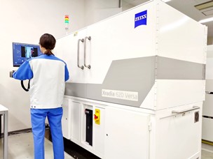
Figure 1. X-ray CT equipment.
Features
By irradiating the sample with X-rays, a two-dimensional transmission image of the internal structure of the sample is obtained. X-ray computed tomography (CT) images are also constructed from the data obtained by rotating the sample.
- Non-destructive.
- Reveals the internal structure of sample and defects.
- 3D images with any viewpoint.
- Cross-sectional images at any point.
- Wide X-ray energy range (30 – 160 kV) allows for a wide range of applications from organic materials to inorganic electronic components.
- Specialized stages enable measurements under tensile/compressive load and/or with temperature control.
Application Examples
- Assessment of fibre shape/dimension/orientation in carbon fibre reinforced plastics (CFRP).
- Shape evaluation and defect investigation of electronic components such as semiconductor devices and their packaging.
- Examination of voids and foreign substances in welded aluminium parts.
- Evaluation of void shape and dimensions in urethane materials.
- Granule shape, dimensions, and fill ratio evaluation in drug capsules.
- Internal shape evaluation of drug tablets.
- Confirmation of regions of interest for physical analysis, such as identification of cross-sectional machining positions.
- Evaluation of product workmanship and quality assurance.
Principle
The sample is irradiated with X-rays and two-dimensional transmission images of the internal structure are acquired while rotating the sample. A 3D X-ray computed tomography (CT) image is constructed from the 2D images, which can be examined from any angle by changing the viewpoint in the browser software. Arbitrary cross sections can also be viewed.
Equipment configuration
X-ray tube, sample manipulator, detector, main processor and image processing software.
Data example
Defective transistor
Carbon fibre reinforced plastics (CFRP)
Data delivery formats
| PDF file |
Single-capture image data. |
| m3d file |
3D X-ray CT image viewer by MST original software (Only Japanese version of the software is available). |
| MP4 file |
Customized animated rotational data (extra charge). |
| Excel file |
Topological and size distribution analysis (separate charge). |
*Other formats are also available (extra charge). Please contact us for details.
Specifications
Items for enquiries
- Purpose and content of measurement
- Sample information
- Number of samples, shape (dimensions), and material composition.
- Type of sample (bulk or thin-film); layer structure and film thickness (if applicable).
- Expected sample delivery date.
- Handling instructions.
- Preferred due date for preliminary analysis report.
- Due date for delivery of final report.
Caution
- Ceramics and some other materials may undergo color change due to X-ray irradiation.
- CT images of samples containing high-density substances, such as heavy metals, tungsten, or barium, may prove difficult to acquire because X-ray transmittance is low.
- If the sample size is larger than the X-ray field, a clear CT image may not be obtained.
- in-situ mechanical or thermal stress is limited in terms of sample shape/size because special stages are used. Contact us for details.